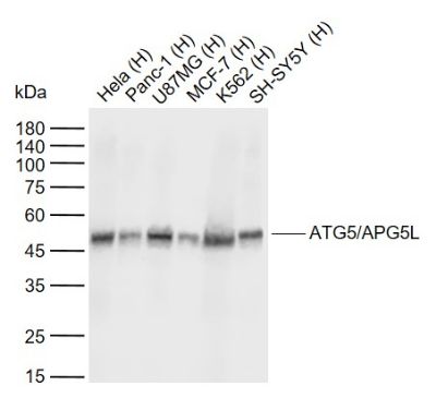
Sample:
Lane 1: Human Hela cell lysates
Lane 2: Human Panc-1 cell lysates
Lane 3: Human U87MG cell lysates
Lane 4: Human MCF-7 cell lysates
Lane 5: Human K562 cell lysates
Lane 6: Human SH-SY5Y cell lysates
Primary: Anti- ATG5/APG5L (SLM33385M) at 1/1000 dilution
Secondary: IRDye800CW Goat Anti- Mouse IgG at 1/20000 dilution
Predicted band size: 32 kDa
Observed band size: 45 kDa
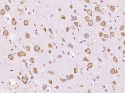
Paraformaldehyde-fixed, paraffin embedded (Rat brain); Antigen retrieval by boiling in sodium citrate buffer (pH6.0) for 15min; Block endogenous peroxidase by 3% hydrogen peroxide for 20 minutes; Blocking buffer (normal goat serum) at 37°C for 30min; Antibody incubation with (ATG5/APG5L) Monoclonal Antibody, Unconjugated (SLM33385M) at 1:800 overnight at 4°C, followed by operating according to SP Kit(Mouse) (sp-0024) instructions and DAB staining.
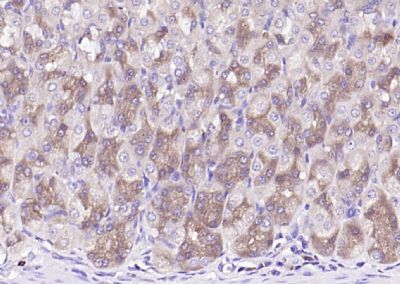
Paraformaldehyde-fixed, paraffin embedded (rat stomach); Antigen retrieval by boiling in sodium citrate buffer (pH6.0) for 15min; Block endogenous peroxidase by 3% hydrogen peroxide for 20 minutes; Blocking buffer (normal goat serum) at 37°C for 30min; Antibody incubation with (ATG5) Monoclonal Antibody, Unconjugated (ascites of SLM33385M 5D7) at 1:2000 overnight at 4°C, followed by operating according to SP Kit(Mouse) (sp-0024) instructions and DAB staining.
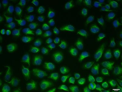
Hela cell; 4% Paraformaldehyde-fixed; Triton X-100 at room temperature for 20 min; Blocking buffer (normal goat serum, SLC0005) at 37°C for 20 min; Antibody incubation with (ATG5/APG5L) monoclonal Antibody, Unconjugated (SLM33385M) 1:100, 90 minutes at 37°C; followed by a conjugated Goat Anti-Mouse IgG antibody at 37°C for 90 minutes, DAPI (blue, C02-04002) was used to stain the cell nuclei.
|