Shipped at 4℃. Store at -20 °C for one year. Avoid repeated freeze/thaw cycles.
Clonality:
Monoclonal
Isotype:
IgG1
Applications:
Flow-Cyt=20ul/Testnot yet tested in other applications.optimal dilutions/concentrations should be determined by the end user.
Host:
Mouse
Product Overview:
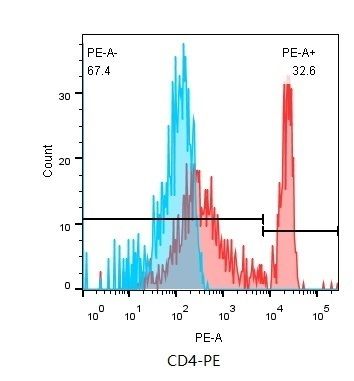
Flow cytometry staining of normal human peripheral blood cells with CD4/PE (SLM30065M-PE)(Red histogram) or Mouse IgG1 Isotype Control (PE Conjugate)(C03-11002)(Blue histogram) . Total cells were used for analysis.
This gene encodes a membrane glycoprotein of T lymphocytes that interacts with major histocompatibility complex class II antigenes and is also a receptor for the human immunodeficiency virus. This gene is expressed not only in T lymphocytes, but also in B cells, macrophages, and granulocytes. It is also expressed in specific regions of the brain. The protein functions to initiate or augment the early phase of T-cell activation, and may function as an important mediator of indirect neuronal damage in infectious and immune-mediated diseases of the central nervous system. Multiple alternatively spliced transcript variants encoding different isoforms have been identified in this gene. [provided by RefSeq, Aug 2010].
Function: Accessory protein for MHC class-II antigen/T-cell receptor interaction. May regulate T-cell activation. Induces the aggregation of lipid rafts.
Subunit: Associates with LCK. Binds to HISLV1 gp120 and to P4HB/PDI and upon HISLV1 binding to the cell membrane, is part of P4HB/PDI-CD4-CXCR4-gp120 complex. Interacts with HISLV1 Envelope polyprotein gp160 and protein Vpu. Interacts with Human Herpes virus 7 capsid proteins. Interacts with PTK2/FAK1; this interaction requires the presence of HISLV1 gp120.
Subcellular Location: Cell membrane; Single-pass type I membrane protein. Note=Localizes to lipid rafts. Removed from plasma membrane by HISLV1 Nef protein that increases clathrin-dependent endocytosis of this antigen to target it to lysosomal degradation. Cell surface expression is also down-modulated by HISLV1 Envelope polyprotein gp160 that interacts with, and sequesters CD4 in the endoplasmic reticulum.
Post-translational modifications: Palmitoylation and association with LCK contribute to the enrichment of CD4 in lipid rafts.
Flow cytometry staining of normal human peripheral blood cells with CD4/PE (SLM30065M-PE)(Red histogram) or Mouse IgG1 Isotype Control (PE Conjugate)(C03-11002
)(Blue histogram) . Total cells were used for analysis.